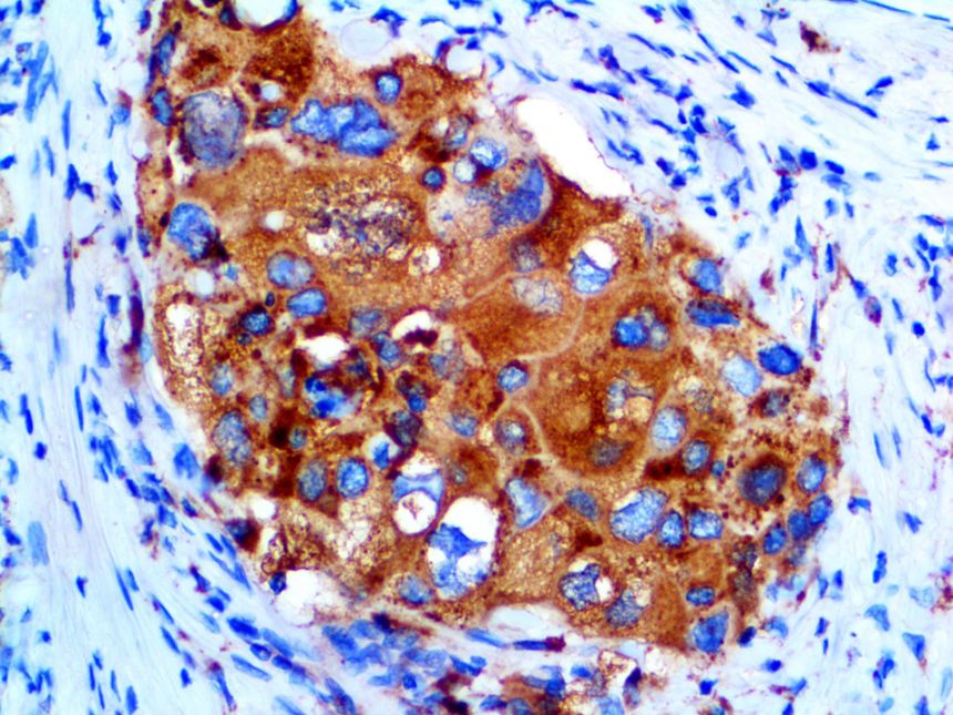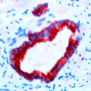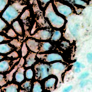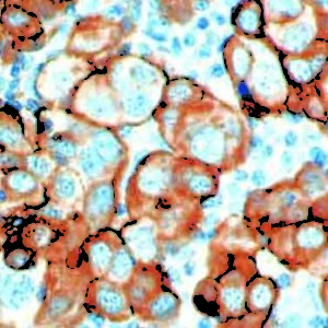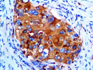
IHC of Napsin on an FFPE Lung Adenocarcinoma Tissue
| Intended Use | For In Vitro Diagnostic Use | |||||||||||||||||||||||||||||||||||
| Summary and Explanation | The activation peptides of aspartic proteinases play a role as inhibitors of the active site. These peptide segments, or pro-parts, are deemed important for correct folding, targeting, and control of the activation of aspartic proteinase zymogens. The pronapsin A gene is expressed predominantly in lung and kidney. Its translation product is predicted to be a fully functional glycosylated aspartic proteinase precursor containg an RGD motif and an addition 18 residues at its C-terminus. In normal tissue, anti-Napsin A labels type II pneumocytes in adult lung and epithelial cells in kidney tissues. In abnormal tissues, Napsin A is a useful marker for lung adenocarcinoma. | |||||||||||||||||||||||||||||||||||
| Antibody Type | Rabbit Monoclonal | Clone | EP205 | |||||||||||||||||||||||||||||||||
| Isotype | IgG | Reactivity | Paraffin, Frozen | |||||||||||||||||||||||||||||||||
| Localization | Cytoplasmic | Control | Kidney, Lung, Lung Carcinoma, Renal Cell Carcinoma | |||||||||||||||||||||||||||||||||
| Presentation | Napsin A is a rabbit monoclonal antibody derived from cell culture supernatant that is concentrated, dialyzed, filter sterilized and diluted in buffer pH 7.5, containing BSA and sodium azide as a preservative. | |||||||||||||||||||||||||||||||||||
| Availability |
| |||||||||||||||||||||||||||||||||||
| Note: For concentrated antibodies, please centrifuge prior to use to ensure recovery of all product. | ||||||||||||||||||||||||||||||||||||
