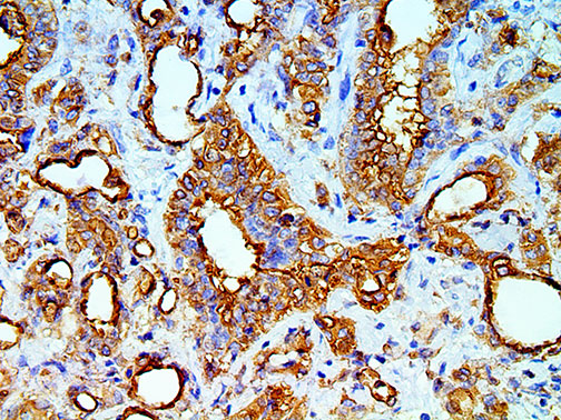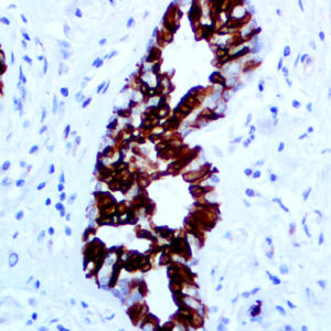
IHC of Mesothelial Cell on a FFPE Mesothelioma Tissue
| Intended Use | For In Vitro Diagnostic Use | |||||||||||||||||||||||||||||||||||
| Summary and Explanation | Mesothelial Cell HBME-1 has shown to label mesothelial cells, both benign and malignant (malignant mesothelioma) and thus has been used in distinguishing mesothelioma from adenocarcinomas of various origins. HBME-1 has also been used to distinguish Thyroid carcinomas (both Follicular and Papillary) from benign thyroid lesions. Mesothelial Cell HBME-1 and MOC-31 have been shown to have a diagnostic efficiency for the distinction between carcinoma and mesothelioma in pleura. HBME-1 staining may be useful for differentiating papillary carcinomas from follicular carcinomas; in papillary lesions it tends to be positive. Several immunohistochemical markers have been used to aid in the diagnosis of follicular-derived lesions of the thyroid (FDLT). HBME-1, ERK, and p16 were found to be more specific for malignancy, whereas CK19 and GAL-3 stained benign lesions with a higher frequency and were not specific for malignant FDLT. A study of thyroid nodules with cytological atypia with strong/diffuse positivity for both HBME-1 and Galectin-3, two well recognized markers of papillary thyroid carcinomas (PTC), represent a starting phenotypic change towards PTC, for which a benign or borderline counterpart has not yet been defined. The expression of HBME-1 and Galectin-3 in some thyroid nodules is related to the presence of cytological atypia suggestive but not diagnostic of PTC. The phenotypic similarity between this subset of thyroid nodules with cytological atypia and PTC is also confirmed by data according to which Galectin-3 and HBME-1 have been found to be highly sensitive for PTC. | |||||||||||||||||||||||||||||||||||
| Antibody Type | Mouse Monoclonal | Clone | HBME-1 | |||||||||||||||||||||||||||||||||
| Isotype | IgGM/K | Reactivity | Paraffin, Frozen | |||||||||||||||||||||||||||||||||
| Localization | Cytoplasmic, Membranous | Control | Breast, Tonsil, Lung, Salivary Gland, TCC, Mesothelioma | |||||||||||||||||||||||||||||||||
| Presentation | Mesothelial Cell is a mouse monoclonal antibody derived from cell culture supernatant that is concentrated, dialyzed, filter sterilized and diluted in buffer pH 7.5, containing BSA and sodium azide as a preservative. | |||||||||||||||||||||||||||||||||||
| Availability |
| |||||||||||||||||||||||||||||||||||
| Note: For concentrated antibodies, please centrifuge prior to use to ensure recovery of all product. | ||||||||||||||||||||||||||||||||||||


