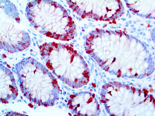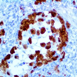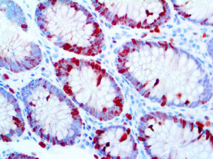
IHC of Ki-67 on an FFPE Colon Tissue
| Intended Use | For In Vitro Diagnostic Use | |||||||||||||||||||||||||||||||||||
| Summary and Explanation | The Ki-67 protein is a cellular marker for proliferation. It is strictly associated with cell proliferation. During the interphase, the Ki-67 antigen can be exclusively detected within the cell nucleus, whereas in mitosis most of the protein is relocated to the surface of the chromosomes. Ki-67 protein is present during all active phases of the cell cycle (G1, S, G2, and mitosis), but is absent from resting cells (G0). Ki-67 is an excellent marker to determine the growth fraction of a given cell population. The fraction of Ki-67-positive tumor cells (the Ki-67 labeling index) is often correlated with the clinical course of cancer. The best-studied examples in this context are Carcinomas of the Prostate and the Breast. | |||||||||||||||||||||||||||||||||||
| Antibody Type | Rabbit Monoclonal | Clone | EP5 | |||||||||||||||||||||||||||||||||
| Isotype | IgG | Reactivity | Paraffin, Frozen | |||||||||||||||||||||||||||||||||
| Localization | Nuclear | Control | Testis, Tonsil, Bone Marrow, Placenta, colon, Tonsil, Fallopian tube, Astrocytoma, Breast Carcinoma, Colon Carcinoma | |||||||||||||||||||||||||||||||||
| Presentation | Ki-67 is a rabbit monoclonal antibody derived from cell culture supernatant that is concentrated, dialyzed, filter sterilized and diluted in buffer pH 7.5, containing BSA and sodium azide as a preservative. | |||||||||||||||||||||||||||||||||||
| Availability |
| |||||||||||||||||||||||||||||||||||
| Note: For concentrated antibodies, please centrifuge prior to use to ensure recovery of all product. | ||||||||||||||||||||||||||||||||||||
| * The Ki-67 antibody, clone EP5, has been manufactured using Epitomics RabMab® technology covered under Patent No.’s 5,675,063 and 7,402,409. | ||||||||||||||||||||||||||||||||||||
