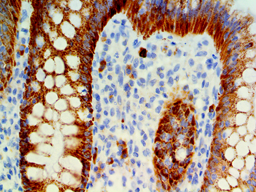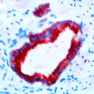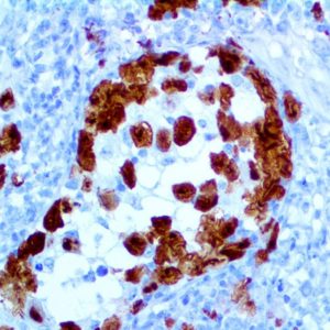
IHC of IMP-3 on an FFPE Colon Tissue
| Intended Use | For In Vitro Diagnostic Use | |||||||||||||||||||||||||||||||||||
| Summary and Explanation | Insulin-like growth factor 2 mRNA-binding protein 3 is a protein that in humans is encoded by the IGF2BP3 gene. The protein encoded by this gene is primarily found in the nucleolus, where it can bind to the 5’ UTR of the insulin-like growth factor II leader 3 mRNA and may repress translation of insulin-like growth factor II during late development. The encoded protein contains several K Homology domains, which are important in RNA binding and are known to be involved in RNA synthesis and metabolism. IMP3 is normally expressed in early embryonic tissues. The IHC of IMP3 may help in the classification of Non-small Cell Lung Carcinomas and Pancreatic Adenocarcinomas as well as subtypes of carcinomas from other organs such Renal Cell Carcinoma, Adenocarcinoma of the Uterine Cervix, Endometrial Carcinoma, Adenocarcinoma of the Esophagus, Malignant Melanoma, Merkel Cell Carcinoma, Urothelial Carcinoma, Neuroendocrine Carcinoma of the Lung, and triple negative breast cancer. Various studies have found that IMP3 is a marker for malignancy and is correlated with increased tumor aggressiveness and reduced overall survival. The diagnosis of Pancreatic Ductal Adenocarcinoma (PDAC) in core needle biopsies can be challenging, and immunohistochemical studies of IMP3 expression have been found to be a potential new marker for the diagnosis of PDAC in core needle biopsies. IMP3 expression for the prognostic evaluation of non-small cell lung carcinomas has been found to exhibit mainly cytoplasmic staining pattern in the NSCLC tissues with a positive rate of IMP3 protein expression of 74.7% in the NSCLC tissues, a significantly higher than the rate of 19.9% in the adjacent non-tumor tissues. In the early- and late-stage NSCLC the disease-free and overall survival rates of the patients with IMP3 expression were significantly lower than those of the patients without IMP3 expression. IMP3 plays a significant role in the progression of NSCLC, and that it may potentially be used as an independent biomarker for prognostic evaluation. IMP3 may be a useful diagnostic marker in the assessment of endometrial cancers and their precursor lesions, particularly when the amount of available tissue material is limited and a concern of type II cancer arises. High frequency of IMP3 expression is present in decidualized endometrial stroma of gestational endometrium and chorionic villi in early pregnancy. | |||||||||||||||||||||||||||||||||||
| Antibody Type | Rabbit Monoclonal | Clone | EP286 | |||||||||||||||||||||||||||||||||
| Isotype | IgG | Reactivity | Paraffin, Frozen | |||||||||||||||||||||||||||||||||
| Localization | Cytoplasmic, Nuclear | Control | Placenta, Tonsil, Ovarian Carcinoma, TCC | |||||||||||||||||||||||||||||||||
| Presentation | IMP-3 is a rabbit monoclonal antibody derived from cell culture supernatant that is concentrated, dialyzed, filter sterilized and diluted in buffer pH 7.5, containing BSA and sodium azide as a preservative. | |||||||||||||||||||||||||||||||||||
| Availability |
| |||||||||||||||||||||||||||||||||||
| Note: For concentrated antibodies, please centrifuge prior to use to ensure recovery of all product. | ||||||||||||||||||||||||||||||||||||


