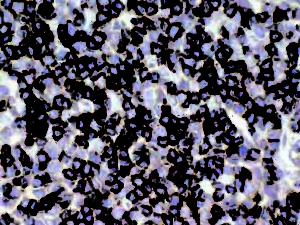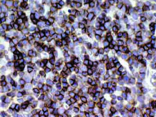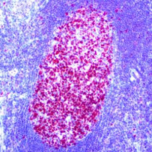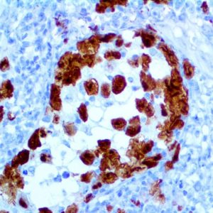
IHC of CD1a on an FFPE Thymus Tissue
| Intended Use | For In Vitro Diagnostic Use | |||||||||||||||||||||||||||||||||||
| Summary and Explanation | CD1 proteins have been demonstrated to restrict T-cell response to non-peptide lipid and glycolipid antigens. At least five CD1 genes (CD1a, b, c, d, and e) have been identified. CD1a belongs to a family of glycoproteins expressed on the surface of various human antigen-presenting cells. In particular, CD1a is a protein of 43 to 49 kDa, and has been shown to be expressed on dendritic cells and cortical thymocytes. Langerhans cells in the skin and some epithelia also express this protein. This antigen is expressed in cells comprising Langerhans Cell Histiocytosis and Langerhans Cell Sarcoma. Anti-CD1a has been used to differentiate various cutaneous Lymphomas (T-cell) from B-cell Lymphomas and Pseudolymphomas. CD1a is also expressed by some malignancies of T-cell lineage and in Histiocytosis X. | |||||||||||||||||||||||||||||||||||
| Antibody Type | Rabbit Monoclonal | Clone | EP80 | |||||||||||||||||||||||||||||||||
| Isotype | IgG | Reactivity | Paraffin, Frozen | |||||||||||||||||||||||||||||||||
| Localization | Cytoplasmic, Membranous | Control | Skin, Thymus, Lymphoblastic Lymphoma | |||||||||||||||||||||||||||||||||
| Presentation | CD1a is a rabbit monoclonal antibody derived from cell culture supernatant that is concentrated, dialyzed, filter sterilized and diluted in buffer pH 7.5, containing BSA and sodium azide as a preservative. | |||||||||||||||||||||||||||||||||||
| Availability |
| |||||||||||||||||||||||||||||||||||
| Note: For concentrated antibodies, please centrifuge prior to use to ensure recovery of all product. | ||||||||||||||||||||||||||||||||||||
| * The CD1a antibody, clone EP80, has been manufactured using Epitomics RabMab® technology covered under Patent No.’s 5,675,063 and 7,402,409. | ||||||||||||||||||||||||||||||||||||


